 The word “microscope” is originated from Greek language words “micro” means small, and “skopion” means to see. In other words, a microscope allows a person to see something too small that can’t be observed with naked eyes. In the scientific world, the instrument of the microscope has brought a revolution that was invented by Zacharias Janssen. Moreover, the discovery of the Cell (a basic block of life) by Robert Hook in 1665 with the help of a microscope was extraordinary.
What Is A Microscope?
The Microscope is a scientific instrument that helps us to see very tiny things (e.g. bacteria) larger and clearer. Moreover, the microscope also helps in measuring dimensions (i.e. length) of microscopic things. Furthermore, microscopes are only helpful in viewing small things (viruses, cells, and crystals) that kept closer to the microscope’s objective lens. They can’t be used to observe something far away.
In biology, microscopes are mostly used to study organisms that are unicellular (e.g. amoeba and euglena) and multicellular organisms. However, microscopes are not limited to biological observations, they are also used in other disciplines such as chemistry and physics. For example, scientists can observe atoms and Molecules under a special and advanced microscope called SEM (Scanning Electron Microscope.
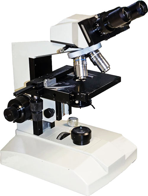
Parts of a Microscope
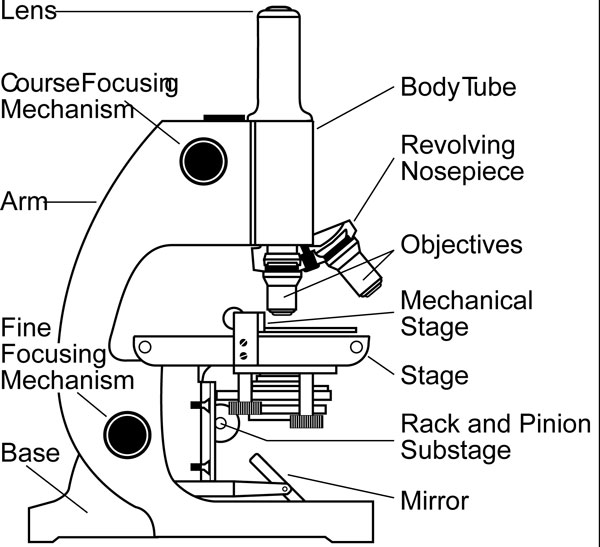 There are various types of microscopes and each type has its own parts. In this section, the parts of a common microscope (optical microscope) are discussed.
- Tube/head – It is the main tube of an optical microscope on with eyepieces and objective lenses are attached.
- Eyepiece/Ocular – It is a group of lenses packed in a container. The sample under observation is seen through eyepieces.
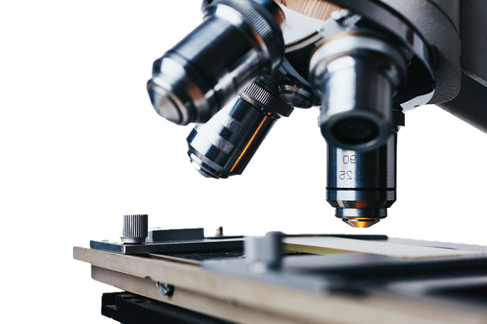
- Objective lenses – These are closest lenses to the sample under observation. It is used to focus and zoom the sample. There are normally 3 objective lenses in a budget class microscope, each objective lens has different magnification.
- Revolving nose piece – small moving disc on which objective lenses are attached. This disc helps in selecting the required objective lens for observing the sample.
- Slide – It is a simple rectangular glass piece used to hold the sample for observation.
- Stage clip – it is a metal clip that is used to hold the slide.
- Condenser – It is used to control the amount of light towards the slide.
- Light source – An LED that is used to produce light that is passed through the condenser.
- Coarse focus – It is a rotating knob used to fine-tune the detail of the sample.
- Fine focus – It is also a rotating knob that is used for focusing the sample.
- Arm – It connects the base to the main tube of a microscope.
- Base – The support of a microscope on which the microscope stands firmly.
Types of Microscopes
 There are many types of microscopes that are in use around the world for various purposes. Some microscopes are useful for cell study while others are good for Soil study. Some of the most important and famous types of microscopes are:
- Simple Microscope – Microscopes that have only one lense are categorized as simple microscopes. In general, magnifying glasses are considered as simple microscopes. These microscopes can have a maximum magnification of 300x. Nowadays, simple microscopes are not used anymore other than magnifying glasses.
- Compound microscope – Compound microscope is better than a simple microscope and consists of more than one lenses. The magnification of these microscopes reaches 1000x to 2000x and gives a much clear view. These microscopes are normally used in biology labs of schools and pathological labs.
- Stereo microscope – Stereo microscope gives a 3D view of the sample under observation. It can also observe objects which are opaque, such as crystals, stones, metals, and jewelry. However, the magnification of these microscopes is very limited. It magnifies objects in a 3D view up to 300 times.
- Confocal microscope – It uses laser light to focus on the sample for increased resolution and sharper image.
- Scanning electron microscope (SEM) – SEM is the modern and one of the most advanced microscopes that magnifies a specimen million times. SEMs don’t require light to focus on the sample. Instead, it uses beams of electrons to get the image of the sample. SEMs are very useful in the field of biology, chemistry, and physics.
- Transmission electron microscope (TEM) – Transmission electron microscope works on the same principals of SEM. TEM differs from SEM only in detection methods of electrons to form an image of the sample. Furthermore, TEM gives a sharper image than SEM but it can observe a much smaller area than the SEM.
History Of Microscopes
 The basic microscope that could be used to magnify objects was first developed by Zacharias Janssen 1590s. After that, the developments continued and the microscope’s resolution and magnification improved. The next leap in the development of the microscope was taken by Robert Hook when he discovered cell 1665. He made and used the microscope that was consisted of 3 lenses. Day-by-day the microscopes have been updated, and nowadays digital microscopes and scanning electron microscope (SEM) are in use.
Interesting Facts
- Galileo Galilei made a better version of the microscope in 1609 along with the development of telescopes.
- Human eyes are capable of seeing objects that are 0.1 mm (100 micrometers) in size under good conditions.
- Sometimes the sample under observation is very transparent and its structure can’t be seen. In this case, the sample is first stained with a color to make its structures visible.